|
Bottom Line: In this paper, we classify the
different types of heterogeneous flow patterns of
microcirculatory abnormalities found during different types of
distributive shock.Analysis of these patterns gave a five class
classification system to define the types of microcirculatory
abnormalities found in different types of distributive shock and
indicated that distributive shock occurs in many other clinical
conditions than just sepsis and septic shock.Functionally,
however, they all cause a distributive defect resulting in
microcirculatory shunting and regional dysoxia.
Affiliation: Department of Physiology, Academic Medical Center,
University of Amsterdam, Amsterdam, The Netherlands.
Abstract: Over 30 years ago Weil and Shubin proposed a
re-classification of shock states and identified hypovolemic,
cardiogenic, obstructive and distributive shock. The first three
categories have in common that they are associated with a fall
in cardiac output. Distributive shock, such as occurs during
sepsis and septic shock, however, is associated with an abnormal
distribution of microvascular (sublingual
microcirculation,clinical
microcirculation,Tongue
microcirculation,lingua
microcirculation) blood flow and metabolic distress
in the presence of normal or even supranormal levels of cardiac
output. This Bench-to-bedside review looks at the recent
insights that have been gained into the nature of distributive
shock. Its pathophysiology can best be described as a
microcirculatory and mitochondrial distress syndrome, where time
and therapy form an integral part of the definition. The
clinical introduction of new microcirculatory imaging
techniques, such as orthogonal polarization spectral and
side-stream dark-field imaging, have allowed direct observation
of the microcirculation(sublingual
microcirculation,clinical
microcirculation,Tongue
microcirculation,lingua
microcirculation) at the bedside. Images of the sublingual
microcirculation during septic shock and resuscitation have
revealed that the distributive defect of blood flow occurs at
the capillary level. In this paper, we classify the different
types of heterogeneous flow patterns of microcirculatory(sublingual
microcirculation,clinical
microcirculation,Tongue
microcirculation,lingua
microcirculation)
abnormalities found during different types of distributive
shock. Analysis of these patterns gave a five class
classification system to define the types of microcirculatory
abnormalities found in different types of distributive shock and
indicated that distributive shock occurs in many other clinical
conditions than just sepsis and septic shock. It is likely that
different mechanisms defined by pathology and treatment underlie
these abnormalities observed in the different classes.
Functionally, however, they all cause a distributive defect
resulting in microcirculatory shunting and regional dysoxia. It
is hoped that this classification system will help in the
identification of mechanisms underlying these abnormalities and
indicate optimal therapies for resuscitating septic and other
types of distributive shock.
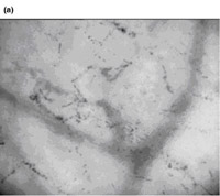
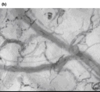
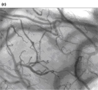
Figure 3: Examples of sidestream dark-field images of
sublingual
microcirculation(clinical
microcirculation,Tongue
microcirculation,lingua
microcirculation) from septic patients with distributive shock.
(a) Image taken from a resuscitated septic patient with a class
I type of microcirculatory abnormality, complete stasis in the
capillaries. (b) An example of a patient with class IV
abnormalities with some capillaries showing stasis and others
showing high flow. (c) Image of a healthy volunteer with
microcirculatory flow in all vessels.


¡¡
Mentions: At the microcirculatory(sublingual
microcirculation,clinical
microcirculation,Tongue
microcirculation,lingua
microcirculation) level, all
classes of abnormalities seen during distributive shock show
normal to hyperdynamic venular flow [8,9]. It is at the
capillary level that the distributive defect is seen, with
heterogeneous perfused capillaries resulting in the shunting of
areas of the microcirculation. Although the classes of capillary
abnormalities we identified may be caused by different
mechanisms, they all have in common a distributive defect caused
by functional shunting of capillaries in the presence of normal
or hyperdynamic venular flow. This is also why we did not make a
distinction between stagnant and stopped flow, as both of these
result in functional shunting. Since microcirculatory(sublingual
microcirculation,clinical
microcirculation,Tongue
microcirculation,lingua
microcirculation)
abnormalities are mainly characterized by a heterogeneous
pattern of flow, we summarized the abnormalities per class in
two main types of capillary flow patterns. This is shown in
cartoon form in Figure 2 as two capillaries below each other,
each with different flow patterns. Venules are depicted as a
single large curved vessel over the capillaries (Fig. 2). In
this way, we identified five classes of sublingual capillary
flow abnormalities (Fig. 2). A Class I abnormality is defined by
all capillaries being stagnant in the presence of normal or
sluggish venular flow (Fig. 3). It is a condition that can be
found in pressure resuscitated septic patients where pressors
have been used excessively to normalize blood pressure [8,9].
Class II microcirculatory flow abnormalities are defined by
empty capillaries next to capillaries with flowing red blood
cells. This decrease of capillary density makes the diffusion
distance between red blood cells in the remaining capillaries
and the tissue cells larger, leading to regional hypoxia [6].
The red blood cells in the remaining capillaries show a high
microcirculatory(sublingual
microcirculation,clinical
microcirculation,Tongue
microcirculation,lingua
microcirculation) hemoglobin saturation, indicating poor oxygen
off-loading associated with the reduction in capillary exchange
surface area [41]. Class II abnormalities were most frequently
found during use of extracorporeal circuits in coronary artery
bypass grafting (CABG) surgery and extracorporeal membrane
oxygenation (ECMO). Class III abnormalities are described by
capillaries with stagnant blood cells next to capillaries with
normal flow. These abnormalities were most frequently observed
in sickle cell patients and critically ill malaria patients, but
also in septic patients. In critically ill malaria patients, who
are often in a coma, strikingly normal hemodynamics are seen in
the presence of high lactate levels. This feature, together with
class III microcirculatory abnormalities, also identifies this
condition as distributive shock. Class IV abnormalities show
hyperdynamic flow patterns in some capillaries next to
capillaries with stagnant cells (Fig. 3). Venules in such cases
frequently also show a hyperdynamic flow profile. This condition
is seen in resuscitated hyperdynamic septic patients. Class V
abnormalities describe the condition where hyperdynamic flow is
seen at all levels of the microcirculation(sublingual
microcirculation,clinical
microcirculation,Tongue
microcirculation,lingua
microcirculation). Blood cells usually
travel so fast that individual cells can not be distinguished
from each other. Metabolic distress seen under such conditions
could be the result of cells moving too fast to off-load their
oxygen, or, that they may originate from other organs or
compartments being shunted [28]. Interestingly, the class V
types of abnormality are also observed in extreme exercise. The
pathogenic nature of class V abnormalities in septic patients
remains to be determined. In Table 1, the diseases observed so
far are listed next to the different classes of microcirculatory
abnormalities seen in Figure 2. They are by no means complete
and it is hoped that this list will continue to expand as more
insight is obtained into the nature of distributive alterations.
Scoring systems developed to quantify such images should greatly
aid this process [42]. Examples of OPS/SDF(sublingual
microcirculation,clinical
microcirculation,Tongue
microcirculation,lingua
microcirculation) movies of each class
of abnormality can be viewed on our web site [43].
http://openi.nlm.nih.gov/ |
|
Introduction: Microcirculation, the small vessels in the
vasculature, embedded within organs, is responsible for the
distribution of blood within tissues, regulation of blood
pressure, delivery of oxygen and other nutrients.
Sidestream
Dark Field (SDF) imaging(sublingual
microcirculation,clinical
microcirculation,Tongue
microcirculation,lingua
microcirculation) is a stroboscopic imaging modality,
allowing assessment of microcirculation. Blood flow is
quantified using Microvascular Flow Index (MFI), perfused vessel
density (PVD), and the proportion of perfused vessels (PPV). The
objectives were to compare the sublingual microcirculation of
pregnant subjects to that of comparable non-pregnant volunteers.
Methodology: The primary outcome is the difference between the
MFI of pregnant versus non-pregnant subjects. Inclusion criteria
include ASA I-II participants in two groups; Pregnant ¨C non-labouring
women with singleton pregnancies, at term gestation,
Non-Pregnant ¨C healthy female volunteers, matched to pregnant
participants for age ¡À 1 year. Participants were excluded with
hypertensive and other cardiovascular disease, obesity,
diabetes, or caffeine intake within 2 hours. The SDF (sublingual
microcirculation,clinical
microcirculation,Tongue
microcirculation,lingua
microcirculation)device was
be applied to the sublingual mucosal surface obtaining steady
images of at least 20 seconds duration. Each participant was be
asked to provide 20 second measurements in five visual fields.
Data Analysis: Video clips were analysed blindly and at random
to prevent coupling between images. The mean MFI values for each
individual was analyzed using paired t-test. Apriori power
calculation was performed for the difference between two
independent means; effect size 1.125, alpha 0.05, power 0.8.
Fourteen subjects per group results in 82% power. To address our
assumptions and potential drop-outs we will increase the group
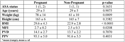
size by 20%. Results: Thirty-seven participants were recruited
(19 pregnant, 18 non-pregnant), a single pregnant participant
was withdrawn because of technical issues. Morphometric(sublingual
microcirculation,clinical
microcirculation,Tongue
microcirculation,lingua
microcirculation)
characteristics are listed in table 1. The PVD and PPV were not
significantly different, while the MFI was significantly higher
in the pregnant group. Conclusion: The microvascular flow index
of pregnant women is higher than a comparable non-pregnant
group, which appears to correlate the physiological changes of
pregnant women. Future projects of this novel imaging technique
should focus on determining the time course of these changes and
the impact of disease process, i.e. preeclampsia and gestational
diabetes on the microvascular and ultimately on the
maternal-fetal unit.

http://soap.org/display_2012_abstract.php?id=S-27 |