|
Abstract
Microcirculation plays a crucial role in the interaction between
blood and tissue both in physiolog¬ical and pathophysiological
states. Orthogonal polarization spectral (OPS) and
Side stream dark field (SDF) imaging
��sidestream dark field (SDF) handheld
imaging device ,Sidestream Dark
Field (SDF) microscopy,Microvascular (blood)
image observation instrument��are relatively new noninvasive methods
that allow the investigation of mucos¬al sites, especially
sublingual area, particularly in critically ill patients. OPS
imaging has been val¬idated against conventional capillary
microscopy, results demonstrated that OPS images provided
similar values for RBC-velocity and capillary diameter as those
measured by conventional capillary microscopy. Despite some
limitations, sublingual microcirculation has been considered as
a possi¬ble surrogate measure for splanchnic blood flow. There
are several areas in human medicine, where observation of
sublingual microcirculatory bed has been carried out �C different
kinds of shock, cardiac arrest, effect of various treatments,
drugs and extreme physiological conditions as well. Early
detection of microvascular abnormality seems to be a key factor
to start early therapeu¬tic intervention in order to reverse
microvascular dysfunction, to maintain efficient microvascular
flow and to contribute to better clinical outcome. In
experimental setting, observing sublingual microcirculation is
an important part of any research focused on the role of
microcirculation dur¬ing various diseases models and to assess
effect of different treatment modalities on microcircula¬tion.
Key words: Microcirculation, sublingual, Orthogonal
polarization spectral imaging (OPS),
Side stream dark field (SDF) imaging
��sidestream dark field (SDF) handheld
imaging device ,Sidestream Dark
Field (SDF) microscopy,Microvascular (blood)
image observation instrument��
Introduction standing of microcirculatory pathology in its
molecular level, especially in critically ill
pa-Microcirculation plays a crucial role in the tients (5, 8,
9). The gold standard for assess¬interaction between blood and
tissue both in ment of microcirculation is intravital
mi¬physiological and pathophysiological states. croscopy (IVM).
However, this technique Analysis of microvascular blood flow
alter-cannot be performed in humans because ations gives a
unique perspective to study there is a need for fluorescent dyes
and tran¬processes at the microscopic level in clinical
sillumination. The size of instrumentation for medicine (1).
Despite the critical role of mi-IVM can be also a limiting
factor for its use crocirculation in numerous diseases includ-in
clinical medicine. For many years, capil¬ing diabetes,
hypertension, sepsis or multiple lary microscopy (capillaroscopy,
nailfold organ failure (2-5), methods for direct visual-videomicroscopy)
has been the only method ization and quantitative assessment of
the for assessment of the human microcircula¬human
microcirculation at the bedside are tion at the microscopic
level in vivo. The use limited (6). The interest in microhemody-of
this technique in humans is limited to eas¬namic monitoring (7)
grows with the under-ily accessible surfaces like the skin,
nailfold,
lip or the bulbar conjunctiva (10). The nail¬fold
microcirculation is extremely sensitive to external temperature
and vasoconstrictive agents (11) and the nailfold
videomicroscopy may not be a reliable indicator of
microcircu¬lation in other parts of the body, particularly in
critically ill patients. Orthogonal polariza¬tion spectral (OPS)
and
Side stream dark field (SDF) imaging
��sidestream dark field (SDF) handheld
imaging device ,Sidestream Dark
Field (SDF) microscopy,Microvascular (blood)
image observation instrument�� represent relatively
new non¬invasive methods that allow the investigation of mucosal
sites, especially sublingual area. There is growing evidence
that relationship between microcirculatory dysfunction and
clinical outcome exists (12, 13). Neverthe¬less, this technique
may be used in experi¬mental settings as well. Access to
sublingual area seems to be very easy, however the key question
is whether this area represents mi¬crocirculation status of
other body tissues or organs; nevertheless, sublingual area
repre¬sents one of the best accessible mucosa sur¬faces in
humans.
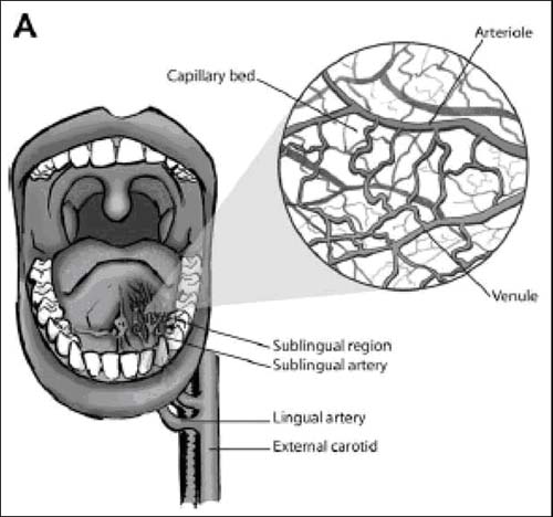
Figure 1: Anatomy of sublin¬gual area (adapted from Klijn, 2008)
Anatomy
The sublingual area is one of the easiest to access areas in
human mucosal surfaces. The major arteries supplying this area
are the ex¬ternal carotid artery, the lingual artery and the
sublingual artery (9, 14-16) (Figure 1). Only limited number of
sublingual arterioles are present, whereas numerous capillaries
(less than 20 um) and venules (20-100 um) are present in this
area (9, 17, 18). External carotid artery contributes to the
perfusion of sublingual mucosa and therefore sublingual
perfusion may reflect cerebral blood flow as well (9). Despite
this fact, only few studies focus on this relationship (19, 20).
Sublin¬gual microcirculation has been considered as a surrogate
measure for splanchnic blood flow, mainly because 1) the tongue
and relat¬ed areas share a common embryogenic ori¬gin with the
gut and 2) the close correlation between sublingual capnometry
and gastric tonometry (21-24).

Imaging techniques
Orthogonal Polarization Spectral Imaging and
Side Stream Dark Field (SDF) Imaging (sidestream dark field (SDF) handheld imaging device ,Sidestream Dark Field (SDF) microscopy,Microvascular (blood) image observation instrument)
Orthogonal Polarization Spectral Imaging (OPS) technology was
invented by Cytomet¬rics, Inc. (Philadelphia, PA, USA) during
the process of developing a videomicroscope able to create high
contrast images of blood in the microcirculation using reflected
light. The original purpose was to develop an in¬strument for
analyzing images of the micro¬circulation using
spectrophotometry in order to compute a complete blood count (CBC)
without removing blood from the body (25,26).
In conventional reflectance imaging (CRI), high-quality image
contrast and detail are limited by multiple surface scattering
and turbidity of the surrounding tissue (25). In OPS imaging,
the main difference from CRI consists in the phenomenon of
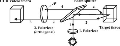
cross-polar¬ization that reduces these effects. As shown in
schematic figure (Figure 2), incident light is linearly
polarized in one plane and pro¬jected through a beam splitter
onto the sub¬ject. Most of the reflected light keeps its
po¬larization and cannot pass through the or¬thogonal polarizer
(analyzer) to form the im¬age. The light that penetrates the
tissue more deeply and undergoes multiple scattering events
becomes depolarized. There is evi¬dence that more than ten
scattering events are necessary to depolarize the light
effec¬tively (27, 28). Hence, only this depolarized scattered
light passing through orthogonal polarizer (analyzer)
effectively back-illumi¬nates absorbing material in the
foreground. A wavelength of the emitted light (548 nm) was
chosen to achieve optimal imaging of the microcirculation
because at this wave¬length oxy- and deoxy-hemoglobin absorb the
light equally. Thus, the blood vessels of the microcirculation
can be visualized by OPS imaging. A detailed description of OPS
imaging technology and further technical im¬provement has been
published previously (26,29). A new device based on OPS
tech¬nology has been developed �C
Side stream dark field (SDF) imaging
��sidestream dark field (SDF) handheld
imaging device ,Sidestream Dark
Field (SDF) microscopy,Microvascular (blood)
image observation instrument��. In this modality a

light guide imaging the microcirculation is surrounded by
light-emitting diodes of a wavelength (530 nm) absorbed by the
hemo¬globin of erythrocytes so that they can be clearly observed
as flowing cells. Covered by a disposable cap the probe is
placed on tissue surfaces. This way of observing the
mi¬crocirculation provides clear images of the capillaries
without blurring (Figure 3) (30, 31). Recent clinical studies of
the human mi¬crocirculation using OPS imaging (Side
stream dark field (SDF) imaging��sidestream
dark field (SDF) handheld imaging device ,Sidestream
Dark Field (SDF) microscopy,Microvascular (blood)
image observation instrument)in various
pathological states have shown a wide spec¬ trum of different
clinical applications with evident impact on diagnosis,
treatment or prognosis assessment. Thus, there is a great effort
to validate OPS imaging for various clinical purposes. The
validation experimen¬tal studies are mostly based on comparison
of IVM and OPS imaging where IVM is sup¬posed to be a gold
standard for main micro¬circulation parameters assessment
(32-34). OPS imaging has been validated especially in animals
(32,35,36) and partly in humans (37). Current knowledge on the
microcircu-

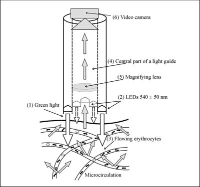
Figure 3: SDF imaging(Side stream dark field (SDF) imaging
,sidestream dark field (SDF) handheld
imaging device ,Sidestream Dark
Field (SDF) microscopy,Microvascular (blood)
image observation instrument), optical scheme. (1) Green light is
emitted by (2) peripheral 540 �� 50 nm light¬emitting diodes (LEDs)
toward tissue arranged in a circle at the end of the light
guide. The microcir¬culation is directly penetrated and
illuminated from the side by green light absorbed by hemoglobin
of erythrocytes which are observed as (3) dark moving cells.
Imaging central part of light guide (4) is optically isolated
from LEDs. A magnifying lens (5) projects the image onto a
camera (6) (adapted from Cerny et al, 2006)
lation is mainly derived from animal studies.
Measurements of the microcirculation in hu¬mans were limited to
easily accessible sur¬faces such as skin and nailfold
capillaries. The basic validation studies in animals have been
performed both on peripheral tissues and solid organs. OPS
imaging techniques (Side stream dark field (SDF) imaging
,sidestream dark field (SDF) handheld
imaging device ,Sidestream Dark
Field (SDF) microscopy,Microvascular (blood)
image observation instrument)has been validated using a highly standard¬ized model of the hamster dorsal skinfold chambre (38).
Four main parameters were measured to validate CYTOSCANTM A/R
against standard fluorescent videomi¬croscopy under normal
conditions and in is¬chemia/reperfusion injury: functional
capil¬lary density (FCD), arteriolar and venular di¬ameter and
venular red blood cell (RBC) ve¬locity. There were not
significant differences between the two techniques for any of
the parameters using Bland-Altman analysis. Similar validation
study in ischemia/reperfu¬sion injury realized using the
CYTOSCAN E-II has confirmed the comparability of OPS imaging and
IVM (38). Functional capillary density is defined as the length
of perfused capillaries per unit area and is given in cm/cm2.
The FCD is a parameter of the tissue perfusion and an indirect
indicator of the oxygen delivery. It is widely used in clinical
studies as semiquantitative method to deter¬mine capillary
density and the proportion of perfused capillaries. OPS imaging
(Side stream dark field (SDF) imaging
,sidestream dark field (SDF) handheld
imaging device ,Sidestream Dark
Field (SDF) microscopy,Microvascular (blood)
image observation instrument)was also validated against IVM in mouse skin flaps and cremaster
muscle preparations. The ve¬locities in straight vessels were
comparable in both methods (39). The dorsal skinfold chamber
model in hamsters was also used to validate OPS imaging under
conditions of hemodilution with a wide range of hemat¬ocrit (Hct)
(38). Bland-Altman analysis of the vessel diameter and FCD
showed good agreement between OPS imaging technique and IVM at a
wide range of Hct. OPS imag¬ing has been validated against IVM
on solid organs in rats, the model of ischemia/reper¬fusion
injury of the rat liver has been used for the assessment of
hepatic microcirculation applying both techniques (40). There
was significant agreement for data obtained from both methods;
correlation parameters for si¬
nusoidal perfusion rate, vessel diameter and venular RBC
velocity were significant. The pancreatic microcirculation has
also been under investigation using OPS imaging (Side stream dark field (SDF) imaging
,sidestream dark field (SDF) handheld
imaging device ,Sidestream Dark
Field (SDF) microscopy,Microvascular (blood)
image observation instrument,)and IVM (41).
Absolute values of the pancreatic functional capillary density
were not signifi¬cantly different between the two methods.
Bland-Altman analyses confirmed good agreement between OPS
imaging and IVM. Thus, OPS imaging is a suitable tool for
quantitative assessment of pancreatic capil¬lary perfusion
during baseline conditions. A murine model of inflammatory bowel
dis¬ease was applied to validate OPS imaging against IVM for the
visualization of colon mi¬crocirculation (42). Postcapillary
venular di¬ameter, venular RBC velocity and FCD were analyzed.
All parameters correlated signifi¬cantly between the both
methods. The as¬sessment of antivascular tumour treatment using
OPS imaging and IVM showed excel¬lent correlation in FCD,
diameter of mi¬crovessels and RBC velocity between both
techniques (35). Validation studies in hu¬mans are limited to
easily accessible sur¬faces; fluorescent intravital microscopy
is excluded because of need to use fluorescent dye. Thus, OPS
imaging has been validated against conventional capillary
microscopy in nailfold skin at rest and after venous occlu¬sion
in healthy volunteers (37). Results demonstrated that OPS images
provided sim¬ilar values for RBC-velocity and capillary
di¬ameter as those measured by conventional capillary
microscopy.
Technical limitations
Despite further development and technical improvement in OPS and
SDF imaging (Side stream dark field (SDF) imaging
,sidestream dark field (SDF) handheld
imaging device ,Sidestream Dark
Field (SDF) microscopy,Microvascular (blood)
image observation instrument)de¬vices (CYTOSCANTM, MicroScanTM
www.mi¬crovisionmedical.com) several limitations re¬main. There
are two main conditions for suc¬cessful OPS/SDF imaging. 1. to
create an im¬age of high quality 2. to evaluate the images as
quantitatively as possible. Three basic technical limitations
have been defined pre¬viously (Lindert et al., n d): undesirable
pres¬sure of the probe affects blood flow, lateral
movement of tissue precludes continuous in¬vestigation of
selected microvascular region, and blood flow velocities above 1
mm/s are difficult to measure, thus information on ar¬teriolar
flow remains hidden. Stabilizing de¬vice which maintains a fixed
distance be¬tween probe and tissue has been developed to
eliminate movement and pressure artefact as much as possible
(43). Image analysis ac¬cording to the principle of spatial
correlation allows extending the range of detectable blood flow
velocities to over 20 mm/s.
The methods for image analysis and quan¬tification in clinical
practise has been report¬ed previously (44), further analysis
improve¬ment using flow scoring system has been published
recently (33). The technology of OPS-SDF imaging (Side stream dark field (SDF) imaging
,sidestream dark field (SDF) handheld
imaging device ,Sidestream Dark
Field (SDF) microscopy,Microvascular (blood)
image observation instrument)has been
incorporated into a small hand-held video-microscope, which can
be used in clinical setting. OPS imaging can assess tissue
perfusion using FCD param¬eter, which is a sensitive parameter
for deter¬mining the status of perfusion to the tissue
and also an indirect measure of oxygen deliv¬ery. The most
easily accessible site in humans is the mouth, where OPS/SDF
technology produces excellent images of the sublingual
microcirculation (Figure 4). However, several limitations should
be acknowledged. Secre¬tions and movement artefacts may impair
im¬age quality. In addition, movement artefacts can spuriously
interrupt flow in some mi¬crovessels. To limit movement
artefacts and to decrease the risk of pressure artefacts, use of
stabilization devices has been proposed (43). Moreover, current
OPS/SDF technology (Side stream dark field (SDF) imaging
,sidestream dark field (SDF) handheld
imaging device ,Sidestream Dark
Field (SDF) microscopy,Microvascular (blood)
image observation instrument)can investigate only tissues covered by a
thin epithelial layer and therefore internal organs are not
accessible, except for perioperative use. Another major problem
with OPS/SDF imaging is the great variability of the vessels
measured. Identical site of interest cannot be examined over
time in contrast to intravital microscopy in animal experiments
(e.g. skin¬fold chamber). Movement artefacts, uncon¬trolled
application pressure, semi quantitative

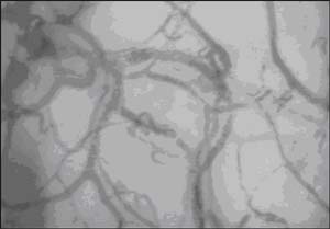
Figure 4: Image of human sublingual microcirculation. SDF
imaging (Side stream dark field (SDF) imaging
,sidestream dark field (SDF) handheld
imaging device ,Sidestream Dark
Field (SDF) microscopy,Microvascular (blood)
image observation instrument)of sublingual area in a healthy volunteer (male, 48
years), captured by MicroScan Video Microscope System
......and so on |